Minimal invasive surgery has been used in gynecology for many years and has become a field of gynecological oncology after the 1990s. Pelvic and para-aortic lymphadenectomy was first reported laparoscopically in endometrium cancer in 1992.1 Over the years, gynecological oncology surgery has become safe to be performed laparoscopically with newly developing energy modalities. Laparoscopic hysterectomy is a surgical operation that has a place in cervical, endometrial and ovarian cancers. The duration of the operation after the laparoscopic surgery is longer than the open surgery, but less pain, shorter hospital stay, and faster return of bowel functions occur after surgery.CLASSIFICATION
In laparoscopic hysterectomy, by definition, the uterine arteries are ligated laparoscopically. Vaginal mucosa can be closed vaginally. In laparoscopic-assisted vaginal hysterectomy described in 1989, round ligaments and adnexal vessels are ligated laparoscopically; Vaginal hysterectomy is performed so that the uterine arteries are connected vaginally. In total laparoscopic hysterectomy, all intraperitoneal ligaments and vascular pedicles are ligated laparoscopically and vaginal surgery is not performed. Laparoscopic hysterectomy is classified as shown in Table 97-1.2

Patient Selection
Appropriate patient selection is a very important issue in laparoscopic hysterectomy. In making this decision, the patient’s age, previous surgical initiations, and body weight should be considered. Obesity is not a stand alone contraindication for laparoscopic surgery.3-5 In obese patients, problems may be encountered in the first entry to the abdomen, excluding the intestines in the abdomen and maintaining a long-term Trendelenburg position. However, less wound infections occur with laparoscopic hysterectomy in obese patients.3,4
Although there are applications that vary between medical centers, the basic steps of laparoscopic hysterectomy described in the literature are the same.6 Absolute contraindications for laparoscopic intervention in terms of anesthesia are shock state, increased intracranial pressure, severe myopia or retinal detachment. A history of bullous emphysema or spontaneous pneumothorax constitutes a relative contraindication.7
In gynecological oncology surgery, the uterus should be taken out of the abdomen without fragmentation. Therefore, the size of the uterus is important in making the decision for laparoscopic hysterectomy. Laparoscopic hysterectomy should not be performed in cases with a lower uterine segment larger than 8 cm, since lateral dissection will be difficult and the uterus cannot be removed vaginally without morselization.8
Mechanical bowel preparation, appropriate antibiotic and low molecular weight heparin prophylaxis should be performed before the surgical operation. The patient should be given detailed information about the surgical procedure, explained that there is a possibility of conversion to open surgery, and written informed consent of the patient should be obtained.

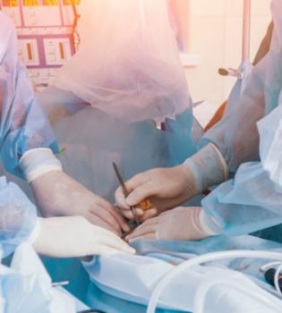
Surgical Technique
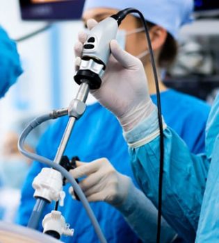
Laparoscopic surgery is a method that uses many surgical instruments and requires the surgical team to work in harmony. Camera, light source, insufflation device, hand manipulation tools, suture tools, intraabdominal fluid delivery and removal system, bipolar electrosurgical systems and uterine manipulator should be reviewed before the operation.
The greatest development in laparoscopic surgery is the development of bipolar electrosurgery systems that provide reliable hemostasis. For coagulation and cutting, ‘Harmonic shears’ (Ethicon, Endo-surgery, USA) 5 mm version and “LigaSure” (Valleylab, USA) 5 and 10 mm models and EnSeal 5 mm (SurgRx, Redwood City, CA) system can be used. 5 mm diameter LigaSure and EnSeal are effective and safe for occlusion of vessels below 7 mm. While the thermal spread in bipolar coagulation can be up to 13 mm, the thermal spread is low in these new bipolar energy systems.9 In electrocauterization, the electrical power is directly converted into heat, while in ultrasonic coagulation devices, ultrasonic sound waves are converted into localized microvibrations in the tissue where the electrical power is applied, and these vibrations create transection and coagulation at lower temperatures; provides occlusion of vessels up to 5 mm. No electrical current passes through the patient. In long-term use, very high temperature values occur at the active and inactive tip. The risky situation is that the thermal damage cannot be seen macroscopically during surgery. Next to important organs such as the ureter, should be worked at low levels (power level 3), and on other organs, should not be worked over power level 4 and should not be applied for longer than 5 seconds. Care should be taken in the tissue around the tip which is active and inactive in use for more than 5 seconds at maximum power and 10 seconds at medium power, and blunt dissection should not be done as the tip will be hot after long-term application.10 After using ultrasonic coagulation scissors, bio-aerosol substances that can be taken through the respiratory tract are formed. Special systems should be established to evacuate the smoke in the abdomen, and contact with potentially infectious blood and blood products and bio-aerosols should be prevented with filter masks.11
After endotracheal anesthesia, the patient is placed on the table in the dorsal lithotomy position. Both arms are placed next to the patient. Gastric decompression is provided with an orogastric or nasogastric tube. Care should be taken to avoid peroneal nerve compression while placing the legs on the table.
In the upright Trendelenburg position, shoulder supports are carefully placed to prevent the patient from slipping. Brachial plexus injury due to shoulder support has been reported in the upright Trendelenburg position with the arms abducted.12 In the upright Trendelenburg position, central venous pressure increases, lung volume decreases, airway ventilation pressure increases, intracranial and intraocular pressures increase. In cases of prolonged hypotension and excessive fluid administration during surgery, intraocular circulation may be impaired due to the upright Trendelenburg position. In addition, venous distension develops in the upper body, tongue and face in the upright Trendelenburg position. Respiratory failure due to laryngeal edema after extubation has been reported.13
If the patient does not have iodine allergy or thyroid disease, it is stained with povidene-iodine complex and covered appropriately. A sterile Foley catheter is inserted into the urethra. Different uterine manipulators can be used for uterine manipulation. An example of uterine manipulators with many different models is the RUMI manipulator and Koh colpotomizer (Coopersurgical, USA) system. The system has four parts: the tip that goes into the uterus with a balloon inside, the metal part that goes through the cervix and inserts into the fornix, the colpo-pneumo-occluder that inflates in the vagina, and the hand-held part that provides the uterus version. The tip that enters the uterus and the colpo-pneumo-occluder are available for single use. The tip, which enters the uterus and has a balloon inside, has three lengths of 6 cm, 8 cm and 10 cm. The metal piece that passes into the cervix and places in the fornix has three dimensions of 3 cm, 3.5 cm and 4 cm. After the cervix being held with a single toothed instrument, the tip with appropriate length is determined by hysterometry. The appropriate tip and the metal piece to be placed in the fornix are selected and the system is placed in the uterus. In this system, there are two balloons that are inflated separately in the uterus and in the vagina. The balloon in the uterus is inflated with 2 cc and the balloon in the vagina is inflated with 60 mL of fluid. Anteversion and retroversion of the uterus are provided by turning the handpiece clockwise and counterclockwise. The balloon in the vagina prevents the loss of pneumoperiotonerum after colpotomy. The metal piece placed in the fornix facilitates the identification of the vaginal fornix during colpotomy.14
The surgical team consists of two surgeons, the camera assistant, the assistant who manipulates the uterus vaginally, and the surgical nurse. Symphysis pubis, iliac crest and umbilicus constitute important structures for port placement. Before inserting the Veress needle, it should be ensured that the stomach and bladder are empty and the patient should be placed in a neutral position on the table. A 10 mm incision is made into the navel. The abdominal wall is lifted by holding the navel on both sides with tissue and laundry forceps, and the Veress needle, which is connected to the insufflator and whose gas flow is open, is entered vertically into the abdomen at a 90° angle. The tip of the Veress needle should be advanced 2 cm past the abdominal wall. The tip of the Veress needle is not moved in the abdomen. The fact that the intra-abdominal pressure is below 10 mm after the Veress needle is inserted indicates that the abdomen has been entered. The primary port through the navel is placed when the intra-abdominal pressure is 25 mm Hg. The primary port is inserted into the abdominal wall at a 90° right angle and is not pushed further when its tip crosses the abdominal wall. When an intra-abdominal image is obtained with the primary port, the entry site and 360° entire intra-abdominal region are checked. In cases with previous abdominal surgery with a Veress needle insertion site, Palmer’s point can be used in the upper left quadrant. During surgery, the abdominal pressure should be kept at 12-15 mm Hg. When placing the right and left lower quadrant ports, the inferior epigastric vessels located in the lateral umbilical fold should be under optical observation. A 10 mm port can be placed on the symphysis pubis in the suprapubic region. Port locations may vary depending on the planned surgical procedure and the surgeon’s preference (Figure 97-1).
The patient is placed in the upright Tredelenburg position. The intestines are mobilized to the upper abdomen. In this period, a decision should be made whether the surgery can be performed laparoscopically.
The presence of ureters prior to hysterectomy allows the surgery to be performed more safely. By placing the uterus in anteversion position, the ureters can be observed intraperitoneally where they enter the pelvis. Using ultrasonic coagulation scissors, bipolar electrocoagulation systems or bipolar cauterization, the full layer of the round ligament is grasped and separated. Ureteral dissection can be performed by entering the retroperitoneal area with an incision made parallel to the lateral part of the infundibulopelvic ligament by having the round ligament on traction. The window under the infundibulopelvic ligament is opened and isolated. The vascular pedicle of the infundibulopelvic ligament is cut using ultrasonic coagulation scissors, bipolar electrosurgical systems, or bipolar coagulation after visualization of the ureter. If hysterectomy is to be performed so that the adnexa is preserved, the utero-ovarian ligament vascular pedicle is cut and separated using the same energy methods. The bladder is separated from the lower uterine segment by making an incision along the vesicouterine fold in the anterior leaflet of the broad ligament. Bladder flap should be formed carefully and thermal damage should be minimized no matter which energy method is used. By means of sharp and blunt dissection, the bladder is separated from the lower uterine segment, cervix and upper vagina.
The next step is to expose the uterine vessels. The peritoneum of the broad ligament is opened and the uterine vessels are isolated. The relationship between ureter and uterine vessels is reviewed. Uterine arteries are cut using ultrasonic coagulation scissors, bipolar electrosurgical systems, or bipolar coagulation cauterization. All three methods have no superiority over each otherr.6 Ligation of the uterine arteries can also be done by means of intracorporeal suture. By placing the uterus in anteflexion, the uterosacral ligaments are separated by means of ultrasonic coagulation scissors, bipolar electrosurgical systems, or bipolar cauterization. During this procedure, damage to the rectum and ureter should be avoided. An anterior colpotomy is made by finding the vaginal-cervical junction and the incision is widened posteriorly. Colpotomy can be performed by means of the active tip of harmonic scissors or by means of monopolar cauterization. If the active tip of the harmonic scissors hits the metal manipulator part in the fornix, there is a possibility that the active tip will breakdown. By holding the cervix with a single-toothed instrument, it is removed from the abdomen by means of the manipulator. Pneumoperitoneum is restored by placing a tampon of appropriate size in a glove into the vagina. The top of the vagina is closed separately with deep sutures that include the uterosacral ligament intracorporeally. Minimal thermal energy should be used at the top of the vagina. The tip of the monopolar instrument used during colpotomy and separation of the uterosacral ligament, and, the tip and the distal 7 cm portion of the ultrasonic coagulation scissors may become very hot; contact with the bladder, ureters and intestines should be avoided and instruments used as energy sources should be carefully moved in the abdomen under continuous optical observation. Bleeding is controlled by washing the top of the vagina with liquid. Bipolar coagulation should not be performed close to the ureter, as lateral thermal damage may occur in hemostasis procedures performed with bipolar cauterization. After the surgical operation is completed, the ports are removed under direct observation. Fascia defects with a diameter of 10 mm through port holes and 5 mm fascia defects that are overused or enlarged should be closed with late-dissolving sutures.
In the post-surgery period, patients have less pain and less analgesia requirements. Post-surgical ileus lasts shorter and normal diet can be started sooner. The hospital stay is shorter.6
Complications
There are some complications particular to laparoscopic hysterectomy. In the first entry to the abdomen with the Veress needle, damage to the large vessels and intestines may occur. Veress needle should be inserted into the abdominal wall at a right angle, the needle should exceed the thickness of the abdominal wall by 2 cm at most and should not be moved in the abdomen. Deep inferior epigastric vessels may be damaged at lateral trocar accesses. Laparoscopic suturing of the abdominal wall may be required to stop the bleeding. Intestinal injury may occur at the first port entrance from the umbilicus. In a large case series where a port was placed from the left upper quadrant (Palmer’s point), the prevalence of umbilical adhesions has been reported as:
0.68% in cases without previous surgery
1.6% in those with previous laparoscopic surgery
19.8% in patients with previous Pfannensitiel incision
51.715% in those with a previous midline incision
Separation of the top of the vagina occurs more frequently after laparoscopic hysterectomy compared to abdominal and vaginal routes; In a series of 7286 hysterectomies, it was reported as 4.9% in total laparoscopic hysterectomy, 0.29% in vaginal hysterectomy, and 0.12% in abdominal hysterectomy.16 Appropriate hemostasis should be performed using minimal thermal energy at the top of the vagina. The top of the vagina should be closed with deep sutures that include the uterosacral ligament.
Before starting laparoscopic hysterectomy, ureter tracing should be revealed by intraperitoneal or retroperitoneal method. Energy sources near the ureter should be used very carefully and ureteral contact with hot instrument tips should be avoided. Sigmoid colon damage due to the use of ultrasonic coagulation scissors has been reported.17 Instruments such as monopolar and ultrasonic coagulation scissors, which may have very high temperatures at the tip and uninsulated distal parts, should be moved in the abdomen under continuous optical observation and contact with the bowel, major vessel, ureter and bladder should be avoided.
The risk of bladder damage was found to be significantly higher in laparoscopic hysterectomy compared to vaginal and abdominal hysterectomy.18 Cystoscopy was performed in all 839 cases who underwent hysterectomy for benign reasons, and 2.9% bladder and 1.8% ureter damage were detected, and 75% of this damage was diagnosed only by cystoscopy.
Undetected bladder perforations may occur during laparoscopic hysterectomy. At the end of the surgical operation, in each case, especially if deep sutures were placed on the top of the vagina, it would be useful to see if sutures pass through the bladder and jet urine flow from the ureteral orifices inside the bladder with cystoscopy. Bladder integrity can also be checked by administering methylene blue into the bladder.19
After the surgical operation is completed, fascia defects of 10 mm port sites and overused or enlarged 5 mm port sites should be closed. In a large series of 5300 cases by Nezhat et al. 20, the risk of incisional hernia was reported as 0.2%. In this series, the omentum in 7 cases and the intestine in 4 cases at both 10 mm and 5 mm port sites were herniated. Factors that increase the risk of port site hernia are excessive manipulation of the port hole, stretching of the fascia while removing the material, and failure to close the fascia defect.
The risk of trocar site metastasis can be reduced by the use of an endobag and, if necessary, complete excision of the trocar site. In the large series of advanced cases, the risk of trocar site metastasis was found to be 0.64%. Trocar site metastasis occurs in cases with malignant ascites and peritonitis carcinomatosis.21
Conclusion
The use of laparoscopic hysterectomy is increasing in gynecological oncology cases. The basic steps of this surgical procedure are the same and laparoscopic skills and suturing techniques can be developed with appropriate training programs.22 In cost analysis, the shortening of the hospital stay reduces the cost, but the excessive use of disposable instruments increases the cost.23
References
Childers JM, Surwit EA. Combined laparoscopic and vajinal surgery for the management of two cases of stage I endometrial cancer Gynecol Oncol 1992; 45: 46-51
Olive DL, Parker WH. The AAGL classification system for laproscopic hysterectomy. Classification Committee of the American Association of Gynecologic Laparoscopists J Am Assoc Gynecol Laparosc 2000;7:9-15
Obermair A, Monolitsas TP. Total laparoscopic hysterectomy versus total abdominal hsyterectomy for obese women with endometrial cancer In J Gynecol Cancer 2005;15:319-324
Eltabbakh GH, Shamanki MI. Hysterectomy for obese women with endometrial cancer: Laparoscopy or laparotomy? Gynecol Oncol 2000;78:329-335
Yu CK, Cutner A. Total laparoscopic hysterectomy as a primary surgical treatment for endometrial cancer in morbidly obese women BJOG 2005;112:115-117
Sokol AL, Gren IC. Laparoscopic hysterectomy. Clinical Obstetrics and Gynecology 2009;52:304-312
Gerges FJ, Kanazi GE. Anesthesia for laparoscopy: a review. J Clin Anesth 2006;18:67-78
Kehoe SM, Levine DA, Abu-Rustum NR. Total laparoscopic hysterectomy In: Ed. Covens AL, Kupets R. Laparoscopic surgery for Gynecologic Oncology McGraw Hill 2009;9-27
Newcomb WL, Hope WW, Schmeizer TM. Comparison of blood vessel sealing among new electrosurgical and ultrasonic devices. Surg Endosc 2009;23:90-96
Kim JS, Hattori R, Yamamoto T. How can we safely use ultrasonic laparoscopic coagulating shears? Int J Urol 2010;17:377-381
Ott De, Moss E. Aerosol exposure from an ultrasonically activated (harmonic) device. J Am Assoc Gynecol Laparosc 1998;1:29-37
Querleu D, Leblanc E. Audit of preoperative and early complications of laparoscopic lymph node dissection in 1000 gynecologic cancer patients. Am J Obstet Gynecol 2006;195:1287-1292
Phing SV. Anesthesia for robotic-assisted radical prostatectomy: considerations for laparoscopy in the Trendelenburg position. Anaesth Intensive Care 2007;35:281-285
Varma R, Gupta JK. Laparoscopic entry techniques: Clinical guideline, national survey, and medicolegal ramifications. Surg Endosc 2008;22:2686-2697
Audebert AJ, Gomel V. Role of microlaparoscopy in the diagnosis of peritoneal and visceral adhesions and in the prevention of bowel injury associated with blind trocar insertion. Fertile Sterile 2000;73:631-635
Hur HC, Guido RS, Mansuria SM. Incidence and patient characteristics of vaginal cuff dehishence after different modes of hysterectomies. J Minim Invasive Gynecol 2007;14:311-317
Atwwad JT, Isaacson K. The harmonic scalpel and intraoperative complications. Obstet Gynecol 1996;88:718-720
Johnson N, Barlow D, Lethaby A. Methods of hysterectomy: systematic view and meta-analysis of randomised trials BMJ 2005;330:1478-1420
Ibeanu OA, Chesson RR, Echols KT. Urinary tract injury during hysterectomy based on universal cystoscopy. Obstet Gynecol 2009;113:6-10
Nezhat C, Nezhat F, Seidman DS. Incisional hernias after operative laparoscopy. J Laparoendosc Adv Surg Tech 1997;7:111-115
Abu Rustum NR. Subcutaneous tumor implantation after laparoscopic procedures in women with malignant disease. Obstet Gynecol 2004;103:480-487
Garrett AJ. Total laparoscopic hysterectomy: The Brisbane learning curve ANZOG 2007;47:65-69
Falcone T, Walters MD. Hysterectomy for benign disease Obstet Gynecol 2008;111:753-767